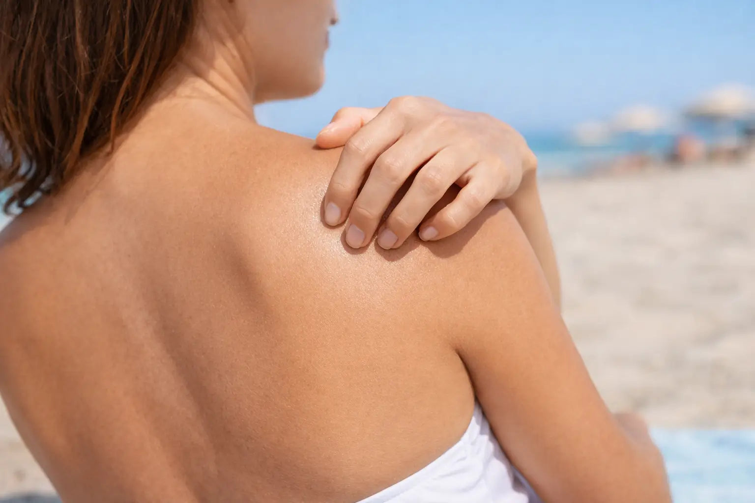
Burning skin without visible lesions is a symptom often ignored by both patients and specialists. The skin appears "normal": no redness, no eruptions, no flaking. Yet, a persistent burning, stinging, warming, or tightness sensation occurs.
Modern dermatology increasingly describes this phenomenon as the result of the interaction of three axes:
- Epidermal barrier (TEWL, lipids, NMF)
- Sensory receptors (TRPV1, TRPA1)
- Neuroinflammation and the skin-brain axis
In this pillar article, we examine the molecular mechanisms, clinical implications, and a research-based management plan.
Clinical Definition: What is Burning Skin Without Lesions?
In the dermatological literature, this symptom is often classified as:
- sensitive skin syndrome (SSS)
- cutaneous dysesthesia
- neurogenic inflammation
According to the definition of the International Forum for the Study of Itch, it is a subjective feeling of discomfort (burning, stinging, pain) that may occur without clinically visible inflammatory changes .
The role of the epidermal barrier and TEWL
What is TEWL?
TEWL (Transepidermal Water Loss) is a key indicator of barrier integrity.
Research has shown that:
- increased TEWL correlates with skin hypersensitivity
- Damage to the barrier increases the penetration of irritants
- Even without visible redness, nerve endings may be activated
(Elias & Wakefield, 2014; Berardesca et al., 2013)
how to rebuild the hydrolipid barrier
Intercellular lipids and ceramides
The epidermal barrier consists of:
- ceramides (approx. 50%)
- cholesterol
- free fatty acids
Reducing the ceramide content leads to:
- TEWL increase
- overactivity of pain receptors
- lowering the tolerance threshold for cosmetics
Studies show that the use of ceramides in skincare reduces subjective burning even in the absence of clinical changes.
Check:

TRPV1 - pain and burning receptor
What is TRPV1?
TRPV1 (Transient Receptor Potential Vanilloid 1) is an ion receptor activated by:
- capsaicin
- high temperature
- acids
- oxidative stress
He is present in:
- keratinocytes
- nerve endings
- inflammatory cells
TRPV1 activation without flushing
Studies have shown that activation of TRPV1 can cause:
- burning sensation
- pinching
- hyperreactivity
without visible inflammation.
(Denda et al., 2001; Caterina et al., 1997)
Excessive use:
- AHA/BHA acids
- retinoids
- alcohol
- mechanical peelings
increases TRPV1 expression.
Neurogenic inflammation
Mechanism
As a result of activation of sensory receptors, the following is released:
- substance P
- CGRP (calcitonin gene-related peptide)
- neurokinin
These mediators:
- dilate vessels
- increase the permeability of the barrier
- increase hypersensitivity
This process may occur subclinically – without erythema.
Skin-brain axis
Stress increases the level of:
- cortisol
- CRH (corticotropin releasing hormone)
CRH acts directly on skin mast cells, increasing neuroinflammation.
(Arck et al., 2006)
Therefore, burning skin without visible changes is often accompanied by:
- chronic stress
- insomnia
- anxiety disorders
Skin Microbiome and Burning
Microbiome dysbiosis leads to:
- reducing lipid production
- increasing immune reactivity
- reducing the tolerance of cosmetics
Staphylococcus epidermidis in physiological amounts supports the barrier, but its disturbances may promote hyperreactivity.
Differential diagnosis
Burning of the skin without lesions may be preceded by:
- rosacea
- shingles (prodromal phase)
- diabetic neuropathy
- vitamin B12 deficiency
- thyroid disorders
Diagnostics recommended for persistent symptoms:
- morphology
- glucose
- B12
- TSH
MMP and accelerated aging in chronic skin burning
What are MMPs?
MMPs (Matrix Metalloproteinases) are enzymes responsible for the degradation of extracellular matrix components—primarily collagen and elastin. Under normal conditions, they participate in skin remodeling. Problems arise when their expression is chronically elevated.
The most important things in the context of aging:
- MMP-1 (collagenase)
- MMP-3
- MMP-9
How does chronic irritation activate MMPs?
Research has shown that:
- oxidative stress
- UV radiation
- activation of TRPV1 receptors
- neuroinflammation
→ lead to an increase in MMP expression by activating the AP-1 (Activator Protein-1) pathway.
(Fisher et al., 1996; Quan et al., 2009)
This means that even if no redness is visible, chronic skin hyperreactivity can accelerate collagen degradation.
TRPV1 and aging
Activation of TRPV1 increases:
- production of pro-inflammatory cytokines
- ROS (reactive oxygen species) levels
- MMP-1 expression
In experimental models, blocking TRPV1 has been shown to reduce UV-induced collagen degradation.
TEWL, microinflammation, and collagen loss
Increased TEWL:
- enhances the penetration of irritants
- activates keratinocytes to produce IL-1α
- triggers the inflammatory cascade
A chronic state of low-grade inflammation (“inflammaging”) promotes degradation of the skin matrix.
Clinical consequences
Prolonged burning of the skin without visible changes can lead to:
- faster wrinkle formation
- loss of elasticity
- flaccidity
- persistent hyperreactivity
Therefore, barrier regeneration is not only a matter of comfort – it is also a matter of aging prevention.
Recovery Plan (4-6 week protocol)
STAGE 1 – Reset (2 weeks)
- acid withdrawal
- no retinoids
- no peeling
- no essential oils
Minimalist routine.
STAGE 2 - Reconstruction of the barrier
Ingredients with proven effectiveness:
- ceramides
- cholesterol
- free fatty acids
- beta-glucan
- panthenol
- niacinamide (≤5%)
- ectoine
Studies show a reduction in TEWL and subjective burning after 2–4 weeks.

STEP 3 - TRPV1 Modulation
Ingredients with modulating potential:
- ectoine
- niacinamide
- oat extract (avenanthramides)
- allantoin
In in vitro studies, they reduced the activation of pain receptors.
STEP 4 - Supporting the skin-brain axis
- improved sleep
- caffeine reduction
- breathing techniques
- omega-3 supplementation
What to avoid?
- denatured alcohol
- menthol
- high LAA concentrations
- excessive exfoliation
- sonic brushes with a damaged barrier
Prognosis
In most cases:
- improvement occurs in 2–6 weeks
- full barrier regeneration up to 8 weeks
- Maintaining the effects requires limiting excessive stimuli
FAQ
Is burning skin without a rash dangerous?
Most often not, but it requires regeneration of the barrier and exclusion of systemic causes.
Can TEWL be measured?
Yes, with a special device – a tewameter – in a clinical setting.
Can stress cause burning skin?
Yes, by activating CRH and mast cells.
Can burning sensation precede rosacea?
Yes, it is often the first symptom.
Does burning skin without a rash mean an allergy?
Not always. Contact allergy usually causes redness, swelling, or a rash. If the burning sensation is not accompanied by visible lesions, the cause is more often a disruption of the epidermal barrier, hyperreactivity of TRPV1 receptors, or neuroinflammation, rather than a classic allergic reaction.
Can burning skin be a symptom of a damaged hydrolipid barrier?
Yes. Increased TEWL exposes nerve endings and increases the penetration of irritants. The skin may sting even without erythema, as the inflammatory process is subclinical. Restoration of intercellular lipids usually alleviates symptoms within 2–4 weeks.
Does TRPV1 activation cause a burning sensation?
Yes. TRPV1 is a receptor that responds to heat, capsaicin, and acids. Its excessive activation increases the influx of calcium ions into nerve cells, causing a burning sensation. This can occur without visible redness, especially in chronic skin irritation.
Does chronic burning accelerate skin aging?
Yes. Prolonged activation of inflammatory pathways and TRPV1 increases the expression of MMPs, which degrade collagen. This process can lead to an accelerated loss of skin firmness and elasticity, even in the absence of obvious inflammatory symptoms.
Can stress cause burning skin?
Yes. Stress increases CRH and cortisol levels, which activate mast cells and increase neuroinflammation. The skin responds with hypersensitivity, burning, and a feeling of heat, often without visible erythema. This mechanism is described as the skin-brain axis.
Can TEWL be lowered with care?
Yes. Cosmetics containing ceramides, cholesterol, and free fatty acids rebuild the lipid structure of the epidermis. Studies have shown a decrease in TEWL after 2–4 weeks of using barrier products, which is associated with reduced burning.
Can burning skin precede rosacea?
Yes. In many patients, the burning sensation and hyperreactivity appear before the onset of flushing. This is an early stage of neurovascular dysregulation and increased sensory receptor reactivity.
Can excessive exfoliation cause burning without any changes?
Yes. Frequent use of acids, retinoids, and peels increases TRPV1 expression and TEWL. Even if the skin isn't red, nerve endings become more reactive, resulting in stinging and burning sensations.
Does the microbiome influence skin burning?
Yes. Dysbiosis reduces lipid production and increases the skin's immune activity. Microbiome imbalances can lower the tolerance threshold and exacerbate neurogenic reactions.
Does barrier regeneration prevent aging?
Yes. Reducing TEWL reduces low-grade inflammation, reduces MMP activation, and supports collagen integrity. This improves elasticity and delays the appearance of wrinkles in the long term.
Bibliography
Arck, P. C. et al. (2006) 'Neuroimmunology of stress: skin takes center stage', Journal of Investigative Dermatology , 126(8), pp. 1697–1704.
Berardesca, E. et al. (2013) 'Sensitive skin: mechanisms and diagnosis', International Journal of Cosmetic Science , 35(1), pp. 2–8.
Caterina, M.J. et al. (1997) 'The capsaicin receptor: a heat-activated ion channel in the pain pathway', Nature , 389, pp. 816–824.
Denda, M. et al. (2001) 'Increased TRPV1 expression in barrier-disrupted skin', Journal of Investigative Dermatology , 117(5), pp. 1309–1314.
Elias, PM & Wakefield, JS (2014) 'Mechanisms of abnormal lamellar body secretion', Journal of Investigative Dermatology , 134, pp. 208–216.
Misery, L. et al. (2014) 'Sensitive skin in Europe', Journal of the European Academy of Dermatology and Venereology , 28(2), pp. 5–9.
Proksch, E., Brandner, J.M. & Jensen, J.M. (2008) 'The skin: an indispensable barrier', Experimental Dermatology , 17(12), pp. 1063–1072.